

The Gold Standard Margin
How SFE-Enabled Fluorescence Imaging Delivers Unprecedented Economic Value
Juan Vegarra
Add paragraph text. Click “Edit Text” to update the font, size and more. To change and reuse text themes, go to Site Styles.
Introduction: The Devastating Cost of the "Invisible" Margin
The goal of cancer surgery is simple: remove all malignant tissue while preserving all healthy, functional tissue. Yet, in many complex oncologic procedures—from head and neck resections to colorectal surgery—the surgeon's best tool for differentiating between cancerous and normal cells is often their naked eye and tactile feel, aided by standard white-light imaging.
This reliance on gross anatomy, rather than molecular pathology, leads to a critical and costly problem: the positive surgical margin.
A positive margin means cancer cells are left behind, necessitating follow-up treatments like repeat surgeries (re-excision), radiation, or chemotherapy. This failure is a financial catastrophe for the healthcare system and a profound burden for the patient.
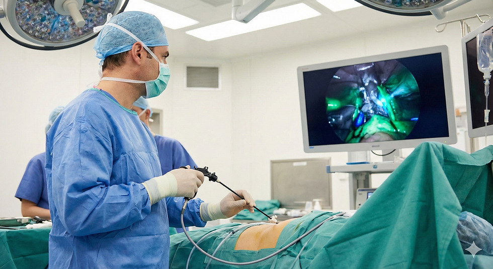
Fluorescence Imaging (FI), or Fluorescence-Guided Surgery (FGS), addresses this failure by making the invisible visible. It uses specialized contrast agents (like Indocyanine Green, or targeted molecular probes) that accumulate in pathological tissue or highlight vital structures. When excited by specific light, these agents glow, creating a real-time, high-contrast map.
The Scanning Fiber Endoscope (SFE) is the ideal platform to deploy this technology endoscopically. Its unique capabilities—ultra-thin, high resolution, and flexible multi-spectral compatibility—allow molecular visualization to be extended into the body's most confined, tortuous, and delicate spaces. The integration of SFE and FI creates a new economic paradigm centered on first-pass success.
Eliminating the Financial Burden of Recurrence and Re-excision
The most direct and substantial economic benefit of SFE-FI is the reduction in positive surgical margins and subsequent recurrence rates.
A. Quantifying the Cost of the Positive Margin
In breast, head and neck, and colorectal cancer surgeries, the rate of positive margins (when cancer cells are found at the edge of the removed tissue) can range from 10% to 30%.
● The Re-operation Cost: A positive margin typically triggers a re-excision surgery. This doubles the surgical cost for the diagnostic episode—incurring a second round of Operating Room (OR) time, anesthesia, hospital stay, and pathology fees. This cost can easily exceed $20,000 to $50,000 per patient, consuming valuable OR capacity and staff resources.
● The Recurrence Cost: When recurrence occurs, it often requires more aggressive, expensive treatments like high-dose radiation or systemic chemotherapy. The lifelong cost of managing recurrent Stage III or IV cancer can be hundreds of thousands of dollars more than the cost of managing the initial Stage I disease.
● The SFE-FI Solution: Studies have shown that Fluorescence-Guided Surgery can drastically reduce the positive margin rate. By allowing the surgeon to see exactly where the tumor ends, SFE-FI ensures a clean margin on the first attempt. For a high-volume center, reducing re-excision rates from 20% to 5% translates into millions of dollars in direct cost avoidance and higher case throughput.
B. Optimizing Tissue Sampling (The Biopsy Dilemma)
In diagnostic endoscopy (e.g., lung, GI), standard white-light visualization can lead to "geographic miss"—biopsying healthy tissue adjacent to the malignancy.
● The Cost of Nondiagnostic Biopsies: A nondiagnostic biopsy requires repeat procedures, which doubles the cost of the diagnostic cycle and delays treatment.
● Molecular Targeting: SFE-FI uses molecular probes that highlight the specific, most metabolically active or dysplastic areas. SFE guides the biopsy tool with molecular precision, ensuring the sample captures malignant tissue. This vastly increases the diagnostic yield of the first biopsy, leading to the "One-and-Done" diagnosis that minimizes patient visits and maximizes the efficiency of the endoscopy suite.
Mitigating Complications: The Financial Value of Anatomical Preservation
Fluorescence imaging is crucial not only for seeing cancer but also for preserving healthy, vital structures like nerves, ureters, and blood vessels. Avoiding iatrogenic injury is a massive component of cost-effective care.
A. Preventing Anastomotic Leakage (The Colorectal Case)
In colorectal surgery, resecting part of the bowel and reconnecting the ends (anastomosis) carries the risk of a leak, often due to poor blood supply (perfusion) at the connection site.
● The Cost of Leakage: An anastomotic leak is a severe, life-threatening complication that requires emergency surgery, often results in a temporary or permanent colostomy, an ICU stay, and can prolong hospitalization by weeks. The cost to manage a leak can exceed $70,000, and this complication directly impacts hospital quality metrics and reimbursement.
● Perfusion Assessment with ICG: SFE-FI, particularly using Indocyanine Green (ICG), provides a real-time, objective assessment of tissue perfusion (blood flow). The SFE's ability to deliver this visualization to complex, deep pelvic anatomy (especially when combined with robotics) ensures the surgeon creates the anastomosis only in well-perfused, viable tissue.
● Cost Avoidance: Economic modeling of ICG-guided surgery suggests a modest but significant cost saving per patient, primarily by reducing the rate of leaks and subsequent re-interventions.
B. Preserving Critical Structures (Nerves and Ducts)
In procedures like prostatectomy, bile duct dissection, or thyroid surgery, accidental nerve or duct injury is costly.
● Liability and Rehabilitation: Injury to the facial nerve, recurrent laryngeal nerve, or bile duct necessitates expensive secondary repairs, long-term physical or speech therapy, and carries substantial legal liability.
● SFE-FI as the Safety Overlay: Specialized fluorescent agents can be developed to illuminate nerves or specific ducts, creating a bright "do not touch" warning sign. SFE’s ultra-high-resolution, stable image provides the precision needed to dissect around these glowing, critical structures, drastically reducing intraoperative damage and long-term functional loss.
Operational Efficiency and Throughput
SFE-FI technology enhances the economic performance of the surgical suite by streamlining decision-making and improving equipment utilization.
A. Reducing Decision Time and Frozen Section Dependence
In cancer surgery, margins are often verified using a frozen section analysis (FSA)—a time-intensive process where tissue is sent to pathology, frozen, cut, stained, and examined while the patient remains anesthetized.
● The Cost of Waiting: FSA adds 20 to 45 minutes of non-productive time to the operation, costing the hospital hundreds to thousands of dollars in OR overhead (staff, anesthesia, utilities) per hour.
● Real-Time Assessment: SFE-FI provides instantaneous margin assessment. The surgeon can see the glowing tumor boundary and confirm clearance in real-time, often rendering the initial, time-consuming FSA unnecessary or allowing the surgeon to precisely target the area of concern for a much quicker analysis. This time savings allows the OR to increase patient throughput by scheduling more procedures per day, directly increasing revenue.
B. Optimizing Equipment TCO (Total Cost of Ownership)
Fluorescence imaging requires specialized optics and light sources. Integrating this capability into the SFE platform offers significant cost advantages over traditional bulky surgical systems.
● SFE’s Miniaturization Advantage: Traditional FGS systems often require large, expensive camera heads and light sources that integrate into standard endoscopes or robotic ports. The SFE, being inherently based on a single fiber, is highly compatible with the precise light delivery needed for fluorescence. This streamlines the hardware, reducing the need for multiple, complex, and fragile filter wheels and bulky optics, which lowers the overall capital cost of the FI system.
● Modular and Disposable Components: If the SFE-FI system utilizes a disposable tip (as in many SFE models), it prevents the catastrophic repair costs associated with damaging the sensitive, integrated fluorescence optics of reusable scopes during high-energy surgical procedures (e.g., laser-ablations).
The SFE-Robotics Multiplier: Accessing High-Value Endoscopic Fields
The highest economic returns are realized when SFE-FI is combined with the precision of Robotics, enabling molecular-guided surgery in previously inaccessible areas.
A. Endoscopic Treatment of High-Value Peripheral Lesions
Many early-stage cancers—such as small, peripheral lung nodules or submucosal GI lesions—are difficult to reach endoscopically with adequate margin control.
● The SFE-FI/Robotic Synergy: The robot provides the stability to keep the scope steady in a moving organ (like the lung), and the SFE-FI provides the microscopic, molecular view needed to confirm an adequate margin.
● Revenue Shift: This enables the endoscopic treatment (e.g., ablation) of tumors that would otherwise require complex, multi-day, high-cost surgical resection. This shift from inpatient surgery to a high-reimbursement, low-complication outpatient procedure is the highest-margin strategy in modern interventional medicine.
B. Personalized Medicine and Monitoring
SFE-FI is an essential component of the emerging personalized medicine market, which is a key growth area for healthcare revenue.
● Theranostics: SFE-FI can be used with specialized molecular probes to not only image the disease but also deliver or track the effectiveness of a targeted drug (a concept called Theranostics). Real-time monitoring of drug uptake via SFE fluorescence ensures that the expensive targeted therapy is working, allowing physicians to stop ineffective treatment early and avoid wasting resources.
● Biomarker Confirmation: SFE-FI can identify specific molecular biomarkers (e.g., EGFR receptors) on the tumor surface. This real-time confirmation of a patient's molecular profile allows for more precise and effective selection of systemic therapies, reducing the cost of using the wrong, ineffective drug.
Conclusion: From Cost Center to Profit Center
The Scanning Fiber Endoscope integrated with Fluorescence Imaging is the technological culmination of decades of research into precision medicine. Its economic value is rooted in its ability to consistently provide a Gold Standard Margin on the first attempt, across a vast range of surgical and diagnostic applications.
For hospital systems, SFE-FI is a crucial investment because it:
Avoids Catastrophic Costs: Significantly reduces re-excision and complication rates (anastomotic leaks, nerve injury), which are the single greatest drains on departmental budgets.
Optimizes Throughput: Eliminates dependence on time-consuming frozen section analysis, maximizing OR utilization.
Drives New Revenue: Unlocks the possibility of high-value, minimally invasive endoscopic procedures in previously inaccessible anatomy, shifting costs from inpatient surgery to high-margin outpatient interventions.
In the era of value-based care, the visibility offered by SFE-FI is not a luxury; it is the most fiscally responsible path forward.
FAQs: SFE in Fluorescence Imaging Economics
1. What is the biggest cost saved by using SFE-FI in cancer surgery?
The largest cost saved is the avoidance of re-excision surgery and subsequent cancer recurrence. Re-excision doubles the cost of the initial surgical procedure (anesthesia, OR time, staffing) and leads to prolonged recovery. Fluorescence imaging, by providing a real-time, molecular view of the tumor margin, drastically increases the chance of achieving a clear margin on the first surgical attempt.
2. How does SFE-FI reduce the risk of a $70,000 anastomotic leak in colorectal surgery?
Anastomotic leaks are often caused by poor blood flow (perfusion) at the surgical connection site. SFE-FI uses the fluorescent dye Indocyanine Green (ICG) to highlight blood flow in real-time. By confirming the tissue has adequate perfusion before the connection is made, SFE-FI helps surgeons avoid high-risk tissue, directly reducing the incidence of leakage and the ensuing, extremely high costs of emergency repair and prolonged critical care.
3. Does fluorescence imaging save money by reducing OR time?
Yes, primarily by minimizing the need for Frozen Section Analysis (FSA). FSA requires the surgical team to wait while tissue samples are processed and analyzed by a pathologist, which can add 20 to 45 minutes of expensive, non-productive time to the procedure. SFE-FI allows the surgeon to visualize the margins in real-time, enabling immediate decision-making and faster overall case completion, thus increasing OR throughput.
4. How does the SFE platform benefit the capital investment in FI technology?
SFE’s miniaturized design simplifies the required optical hardware compared to traditional systems. More importantly, when integrated into a robotic platform, SFE utilizes disposable imaging tips. This protects the core, expensive fluorescence light source and optics from the high risk of damage associated with surgical procedures, leading to a significantly lower Total Cost of Ownership (TCO) and more predictable maintenance costs.
5. What is the long-term economic impact of using SFE-FI in diagnosis?
In diagnostic procedures (like endoscopy or bronchoscopy), SFE-FI increases the diagnostic yield of the first biopsy attempt by guiding the tool to the molecularly active, most malignant part of a lesion. This prevents the costly cycle of nondiagnostic procedures, repeat visits, follow-up imaging, and treatment delays, ultimately ensuring the patient moves into the appropriate, curative care pathway as quickly as possible.