top of page


Intravascular Imaging
Detailed visualization of vascular structures enhances diagnostic precision, helping physicians make accurate assessments. This leads to improved patient outcomes and optimized treatment plans.
Laser Angioscopy: Super vision for interventionalists
3F highly navigable system over-a-wire
True color images of the vascular surface and lumen
Highest resolution intravascular technology
Forward viewing camara
Unsurpassed field or view and massive focal depth (>5cm)
Powerful and “cold” illumination
Artifact-free, intuitive images
Real-time videos
Best in class technology for intravascular diagnosis and image-guided therapy
See clearly.
Diagnose the problem.
Angiographically-occult fibrous cap rupture in carotid artery of patient with cryptogenic stroke.
Ulcerations in non-stenotic carotid artery in patients with recurrent stroke.
See better. Diagnose unequivocally.
 |
Visualization of Erosions (lack of endothelium with red blood cells confirmed with scanning electron microscopy)
See forward.
Treat optimally.
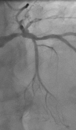
Acute Thrombotic Occlusion | Chronic Calcified Occlusion |
See forward, in real time.
Improve your outcomes.
Image-guided wire advancement in a chronic total occlusion | Super-selective wiring (swine) |
See what is invisible to the eye.
Provide Timely Treatment.
Prevent strokes and heart attacks.


Acute coronary in-stent thrombosis
SFE fluorescence angioscopy visualizing MMP activity in red color in a stented coronary artery
See Deeper.
Know more.
 |  |
Forward looking RGB images of finger | Forward looking OCT images of fingerprint following the yellow line |
In vivo angioscopy
bottom of page